Prof. Anita Arsovska Written Commentary
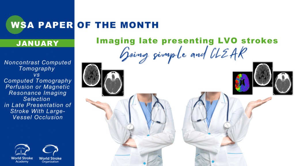
Title: Can non-contrast computed tomography alone be used as an alternative to advanced imaging in selecting patients with late-presenting large-vessel occlusion for mechanical thrombectomy?
Author: Prof. Anita Arsovska, Associate Commissioning Editor WSA
This article is a commentary on the following
Summary:
The CT for Late Endovascular Reperfusion (CLEAR) study was a multicenter cohort study of consecutive patients with proximal anterior circulation stroke undergoing mechanical thrombectomy (MT) in the extended time window, defined as a period from TLSW to arterial puncture between 6 to 24 hours¹. This study was conducted at 15 sites across 5 countries in Europe and North America. The duration of follow-up was 90 days from stroke onset. Clinical outcomes of patients selected for mechanical thrombectomy by non-contrast computed tomography (CT) vs those selected by computed tomography perfusion (CTP) or magnetic resonance imaging (MRI) in the extended time window were compared.There was no difference in 90-day ordinal mRS shift between patients selected by CT vs CTP (adjusted odds ratio [aOR], 0.95 [95% CI, 0.77-1.17]; P = .64) or CT vs MRI (aOR, 0.95 [95% CI, 0.8-1.13]; P = .55). The rates of 90-day functional independence (mRS scores 0-2 vs 3-6) were similar between patients selected by CT vs CTP (aOR, 0.90 [95% CI, 0.7-1.16]; P = .42) but lower in patients selected by MRI than CT (aOR, 0.79 [95% CI, 0.64-0.98]; P = .03). Successful reperfusion was more common in the CT and CTP groups compared with the MRI group (474 [88.9%] and 670 [89.5%] vs 250 [78.9%]; P < .001). No significant differences in symptomatic intracranial hemorrhage (CT, 42 [8.1%]; CTP, 43 [5.8%]; MRI, 15 [4.7%]; P = .11) or 90-day mortality (CT, 125 [23.4%]; CTP, 159 [21.1%]; MRI, 62 [19.5%]; P = .38) were observed. The authors conclude that there were no significant differences in the clinical outcomes of patients selected with non-contrast CT compared with those selected with CTP or MRI. This could potentially widen the indication for treating patients in the extended window using a simpler and more widespread non-contrast CT.
Commentary:
Current guidelines still recommend use of advanced imaging in stroke patients with anterior LVO presenting beyond 6 hours who are candidates for mechanical thrombectomy (refs).The 2018 AHA/ASA Guidelines suggestthat in selected patients with a large vessel occlusion within 6-24 hours from last known normal who would also have been eligible for DAWN or DEFUSE 3, obtaining perfusion imaging (CT-P or MR-P) or an MRI with diffusion-weighted imaging (DWI) sequence is recommended to help determine whether the patient is a candidate for mechanical thrombectomy². Similarly, the 2019 ESO/ESMINT Guidelines advise that in adult patients with anterior circulation LVO-related acute ischaemic stroke presenting beyond 6 hours from time last known well, advanced imaging selection is necessary (with moderate quality of evidence and strong strength of recommendation³).
Kim et al. (2019) aimed to elucidate whether MRI-based selection for EVT is safe and effective within and after a 6-hour time window compared with conventional CTA-based selection⁴.Their study found that MRI-based selection for EVT was not associated with improving functional outcome compared with CT-based selection, but might be better at reducing the risk of SICH, despite the delays in all workflow time metrics.
Bouslama et al. (2021) compared the prediction of post-reperfusion infarct volumes and the clinical outcomes across NCCT e-Stroke software versus RAPID (IschemaView, Menlo Park, CA) computed tomography perfusion measurements⁵.Baseline ischemic core volumes measured by e-Stroke Suite software on NCCT performed similarly to RAPID CTP in estimating post-full reperfusion infarct volumes and functional outcomes for both early- and late-presenting patients. It was concluded that NCCT e-ASPECTS software volumes could be used along CTP to further refine selection for thrombectomy and could also represent a viable alternative in centers where access to advanced imaging is limited.
Jadhav et al. (2021) aimed to compare post-EVT outcomes between patients who underwent pre-randomization basic (non-contrast computed tomography [CT], CT angiography only) versus additional advanced imaging (computed tomography perfusion [CTP] imaging) and to determine the association of performance of pre-randomization CTP imaging with clinical outcomes⁶. They analyzed 1348 patients, 610 (45.3%) of whom underwent CTP pre-randomization. The benefit of EVT compared with best medical management was maintained irrespective of the baseline imaging paradigm. There was no difference between the CTP and non-CTP group (17.1% vs 16.1%) (p=0.57). The 90-day modified Rankin Scale score 0–2 in EVT versus control patients was 46.0% (137/298) in the CTP group versus 28.9% (88/305), whereas 44.1% (162/367) in the without CTP group versus 27.3% (100/366) in the control group. Performance of CTP baseline imaging compared with baseline non-contrast CT and CT angiography only yielded similar rates of good outcome (odds ratio, 1.05 [95% CI, 0.82–1.33], adjusted odds ratio, 1.04, [95% CI, 0.80–1.35]).The authors conclude that rates of good functional outcome were similar among patients in whom CTP was or was not performed, and EVT treatment effect in the 0- to 6-hour time window was similar in patients with and without baseline CTP imaging.
Conclusion:
Based on the latest clinical trials, non-contrast computed tomography alone maybe used as an alternative to advanced imaging in selecting patients with late-presenting large-vessel occlusion for mechanical thrombectomy. More data are needed on specific inclusion and exclusion criteria of potential patients.
References:
- Nguyen TN, Abdalkader M, Nagel S, et al. Noncontrast Computed Tomography vs Computed Tomography Perfusion or Magnetic Resonance Imaging Selection in Late Presentation of Stroke With Large-Vessel Occlusion. JAMA Neurol.Published online November 08, 2021. doi:10.1001/jamaneurol.2021.4082
- Powers WJ, Rabinstein AA, Ackerson T, et al., on behalf of the American Heart Association Stroke Council. 2018 Guidelines for the Early Management of Patients With Acute Ischemic Stroke: A Guideline for Healthcare Professionals From the American Heart Association/American Stroke Association. Stroke2018;49:e46-e110.
- Turc G, Bhogal P, Fischer U, et al. European Stroke Organisation (ESO)—European Society for Minimally Invasive Neurological Therapy (ESMINT) guidelines on mechanical thrombectomy in acute ischaemic stroke endorsed by Stroke Alliance for Europe (SAFE). Eur Stroke J. 2019;4(1):6-12. doi:1177/2396987319832140.
- Kim JT, Cho BH, Choi KH et al. Magnetic Resonance Imaging Versus Computed Tomography Angiography Based Selection for Endovascular Therapy in Patients With Acute Ischemic Stroke. Stroke 2019; 50:365-372. https://doi.org/10.1161/STROKEAHA.118.023173
- Bouslama M, Haussen DC, Rodrigues G, Barreira C, Frankel M, Nogueira RG. Novel selection paradigms for endovascular stroke treatment in the extended time window. J Neurol Neurosurg Psychiatry. 2021;jnnp-2020-325284. doi:1136/jnnp-2020-325284
- Jadhav PA, Goyal M, Ospel J et al. Thrombectomy With and Without Computed Tomography Perfusion Imaging in the Early Time Window: A Pooled Analysis of Patient-Level Data. https://doi.org/10.1161/STROKEAHA.121.034331 2021;0:STROKEAHA.121.034331