The Paper of the Month September
02 Sep 2021Direct to Angiography Suite Without Stopping for Computed Tomography Imaging for Patients With Acute Stroke
Editorial
Author: Dr. Gustavo Saposnik, EiC, WSA
This article is a commentary on the following
“Management is efficiency in climbing the ladder of success; leadership determines whether the ladder is leaning against the right wall. Stephen Covey (American author and businessman)”
Endovascular thrombectomy became standard of care following the completion of five RCTs (and subsequent meta-analyses) in 2015.1-3 Major efforts were devoted to improving patients’ workflow and to reduce time to reperfusion.
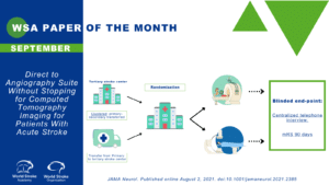
In the present study4, the authors tested the hypothesis of whether stroke patients with presumed large vessel occlusion (LVO) transferred directly to the angiosuite (DTAS) have better process measures and clinical outcomes compared to usual care. Overall, 174 participants with a diagnosis of an acute ischemic stroke with a presumed LVO within 6 hours from symptoms onset were randomly assigned (1:1) to either DTAS (89 patients) or usual care (85 patients). Patients in the usual care group (conventional workflow) received direct transfer to computed tomographic imaging, with usual imaging performed and assessment of the eligibility for EVT.
Findings: Participants randomized to DTAS protocol have a significantly lower median door–to–arterial puncture time (18 minutes [IQR, 15-24 minutes] vs 42 minutes [IQR, 35-51 minutes]; P < .001) and door-to-reperfusion time (57 minutes [IQR, 43-77 minutes] vs 84 minutes [IQR, 63-117 minutes]; P < .001) compared to the standard of care group.
In a shift analysis, participants in the DTAS protocol had two-fold lower degree of disability across the range of the modified Rankin scale (mRS) (adjusted common odds ratio, 2.2; 95% CI, 1.2-4.1; p= 0.009). There were no significant differences in safety outcomes.
The authors concluded that the use of DTAS increased the odds of patients undergoing EVT, had shorter time to reperfusion, decreased hospital workflow time, and improved clinical outcome for stroke patients with LVO admitted within 6 hours after symptom onset.
Commentary: Readers may wonder what the ‘magic’ behind this RCT is, when in fact both groups received brain imaging. What is the difference between having a conventional multidetector brain scan at the CT room versus a flat-CT in the angiosuite?
The combination of several critical factors may explain the positive results of this relatively small trial:
- Improving clinical judgement by using a validated clinical scale: Previous studies showed the benefits of using validated scales for patient selection and stroke prognostication.5, 6 The authors selected candidates with presumed LVO using the Rapid Arterial Occlusion Evaluation [RACE]. Values ≥5 had sensitivity 0.85 and negative predictive value 0.94 for detecting LVO.7
- Overcoming time gaps: transporting and transferring patients in and out the emergency department and CT rooms takes time. There are no diagnostic or therapeutic procedures completed during a transfer or transportation. The lower the number of stations/stops patients undergo, the shorter the time to achieve an effective reperfusion.
- Using a flat-panel CT in the angiosuite: The advantages of flat-panel volume CT over multidetector CT include ultra-high spatial and temporal resolution, dynamic imaging capabilities, and whole-organ coverage in one rotation of the scanner. The disadvantages include a lower contrast resolution and higher radiation.8
- Outcome measures: The authors took the advantage of using continuous variables as process measures and leveraging a shift analysis to determine differences in disability between groups. This strategy increased the likelihood of detecting differences between groups in the transition of health states (e.g. mRS from 4 to 3, 3 to 2, and so on)
- Rational use of resources: The authors used a simplified imaging protocol that was unable to provide thorough parenchymal assessment. This trade-off strategy (prompt but suboptimal imaging in the angiosuite) is appropriate when suspected LVO among patients with an ischemic stroke within 6 hours. An open question relates to whether the same benefit could be achieved for patients beyond 6 hours from symptoms onset.
Limitations of the study include a small sample size, single center and the need of validation in other institutions and countries to ensure the generalizability of results. Another consideration is that 60% of the included patients were initially assessed at a primary stroke center and then transferred to the Val D’Ebron comprehensive stroke center. As a result, most patients assessed at the primary stroke center had a non-contrast CT and an additional CTA as part of the usual care. In other words, over half of participants received usual care prior to the randomization to either DTAS or “continuation of usual care”.
In summary, this RCT provides evidence that combining a selection tool to identify LVO, avoiding delays with intra-hospital transfers, and prompt imaging in the angiosuite improved time to reperfusion and clinical outcomes for patients with an acute ischemic stroke within 6 hours. The authors are to be commended for taking this comprehensive approach that may advance the efficiency in stroke care. If these results are confirmed in other stroke centers, there is a potential for improving the impact of ischemic stroke in our patients and their families.
References
- Powers WJ, Rabinstein AA, Ackerson T, Adeoye OM, Bambakidis NC, Becker K, Biller J, Brown M, Demaerschalk BM, Hoh B, Jauch EC, Kidwell CS, Leslie-Mazwi TM, Ovbiagele B, Scott PA, Sheth KN, Southerland AM, Summers DV and Tirschwell DL. Guidelines for the Early Management of Patients With Acute Ischemic Stroke: 2019 Update to the 2018 Guidelines for the Early Management of Acute Ischemic Stroke: A Guideline for Healthcare Professionals From the American Heart Association/American Stroke Association. Stroke. 2019;50:e344-e418.
- Goyal M, Menon BK, van Zwam WH, Dippel DW, Mitchell PJ, Demchuk AM, Davalos A, Majoie CB, van der Lugt A, de Miquel MA, Donnan GA, Roos YB, Bonafe A, Jahan R, Diener HC, van den Berg LA, Levy EI, Berkhemer OA, Pereira VM, Rempel J, Millan M, Davis SM, Roy D, Thornton J, Roman LS, Ribo M, Beumer D, Stouch B, Brown S, Campbell BC, van Oostenbrugge RJ, Saver JL, Hill MD, Jovin TG and collaborators H. Endovascular thrombectomy after large-vessel ischaemic stroke: a meta-analysis of individual patient data from five randomised trials. Lancet. 2016;387:1723-31.
- Saver JL, Goyal M, van der Lugt A, Menon BK, Majoie CBLM, Dippel DW, Campbell BC, Nogueira RG, Demchuk AM, Tomasello A, Cardona P, Devlin TG, Frei DF, du Mesnil de Rochemont R, Berkhemer OA, Jovin TG, Siddiqui AH, van Zwam WH, Davis SM, Castaño C, Sapkota BL, Fransen PS, Molina C, van Oostenbrugge RJ, Chamorro Á, Lingsma H, Silver FL, Donnan GA, Shuaib A, Brown S, Stouch B, Mitchell PJ, Davalos A, Roos YBWEM, Hill MD and Collaborators ftH. Time to Treatment With Endovascular Thrombectomy and Outcomes From Ischemic Stroke: A Meta-analysis. JAMA. 2016;316:1279-1289.
- Requena M, Olive-Gadea M, Muchada M, Hernandez D, Rubiera M, Boned S, Pinana C, Deck M, Garcia-Tornel A, Diaz-Silva H, Rodriguez-Villatoro N, Juega J, Rodriguez-Luna D, Pagola J, Molina C, Tomasello A and Ribo M. Direct to Angiography Suite Without Stopping for Computed Tomography Imaging for Patients With Acute Stroke: A Randomized Clinical Trial. JAMA Neurol. 2021.
- Saposnik G, Cote R, Mamdani M, Raptis S, Thorpe KE, Fang J, Redelmeier DA and Goldstein LB. JURaSSiC: accuracy of clinician vs risk score prediction of ischemic stroke outcomes. Neurology. 2013;81:448-55.
- Drozdowska BA, Singh S and Quinn TJ. Thinking About the Future: A Review of Prognostic Scales Used in Acute Stroke. Frontiers in Neurology. 2019;10.
- Ossa NPdl, Carrera D, Gorchs M, Querol M, Millán M, Gomis M, Dorado L, López-Cancio E, Hernández-Pérez M, Chicharro V, Escalada X, Jiménez X and Dávalos A. Design and Validation of a Prehospital Stroke Scale to Predict Large Arterial Occlusion. Stroke. 2014;45:87-91.
- Gupta R, Cheung A, Bartling S, Lisauskas J, Grasruck M, Leidecker C, Schmidt B, Flohr T and Brady T. Flat-Panel Volume CT: Fundamental Principles, Technology, and Applications1. Radiographics : a review publication of the Radiological Society of North America, Inc. 2008;28:2009-22.
Author Interview
Marc Ribo, MD, PhD, and Manuel Requena, MD, PhD

1. WHAT DID YOU SET OUT TO STUDY?
With the AngioCat study, we wanted to confirm that for patients suspected to suffer a large vessel occlusion stroke, a Direct-transfer-to. AngioSuite (DTAS) protocol on arrival to the stroke center not only reduces workflow times but is also safe and improves long term outcome.
2. WHY THIS TOPIC?
We started exploring this protocol in 2017 and, as a team acquired sufficient experience to consistently achieve door-to-puncture times below 20 minutes when the protocol is activated. We were convinced that this approach is beneficial for the patient and wanted to build evidence in order to spread DTAS in other large volume endovascular centers.
3. WHAT WERE THE KEY FINDINGS?
Our randomized study confirmed that DTAS can efficiently reduce door to puncture time in around 30 minutes but also that imaging evaluation based on cone beam CT scan in the angio-suite is sufficient to effectively select patients for EVT in the 0-6 hours window. As a result, DTAS leads to a significant shift towards better 90 days disability outcome.
4. SOME PATIENTS WERE TRANSFERRED FROM OTHER CENTERS. CAN YOU CLARIFY HOW MANY PATIENTS HAD A CONVENTIONAL CT/CTA, EVEN IF RANDOMIZED TO DTAS?
In AngioCat 60% of the included patients were initially atended in a primary stroke center and transferred to our comprehensive center. AT primary stroke center all patients had a non-contrast CT and an additional CTA in those centers capable to perform it.
Independently of initial imaging acquired at thE primary stroke center on arrival to the comprehensive stroke center patients were randomized into angiocat to:
- Conventional imaging: repeating CT + CTA
- DTAS: only cone beam CT in the angiosuite + DSA (in case of no ICH observed) to diagnose LVO (none of the patients in this group had received an additional conventional CT scan on admission)
5. CAN YOU PLEASE CLARIFY IF THERE WAS ANY OTHER CONTAMINATION BETWEEN GROUPS?
We did not have any cross-overs between allocated groups.
6. HOW MIGHT THESE RESULTS IMPACT CLINICAL PRACTICE?
Our results should motivate other large volume endovascular capable stroke centres to rethink their admission stroke protocols in order to include the possibility to perform DTAS on patient arrival. A progressive adoption of DTAS in parallel with the acquisition of the necessary team experience and coordination will lead to reduce disability in stroke patients receiving EVT.
7. WHAT SURPRISED YOU MOST?
A deep analysis of our results showed that DTAS might improve outcome not only by reducing workflow times but also by increasing the number of the patients who finaly received EVT. We observed that a higher proportion of patients with a larger infarct core received EVT if evaluated through DTAS as compared to conventional imaging and this may have contributed also to the differences in detected in long term outcome.
8. WHAT’S NEXT FOR THIS RESEARCH?
In other to confirm that our findings are applicable to other networks and settings we recently launched the WE-TRUST study which is designed to confirm the efficacy of DTAS in a multicentric international randomized trial.
9. IS THERE ANYTHING YOU’D LIKE TO ADD?
We’d like to encourage my colleagues to considerate adopting DTAS in their daily practice. DTAS requires a team learning curve and high multilevel coordination, including the pre-alert of patient arrival by prehospital emergency services, in order to achieve substantial time reductions in door-to-puncture times. We would recommend to start with those cases in which all circumstances are favourable (time of the day, team availability…) and progressively expand to a higher number of cases.


